eUnity supports the display of mammography images / exams with modality type as MG or CR and one of the following SOP class UIDs:
- Digital Mammography X-Ray Image Storage for Presentation (1.2.840.10008.5.1.4.1.1.1.2)
- Breast Tomosynthesis Image Storage (1.2.840.10008.5.1.4.1.1.13.1.3)
- Computed Radiography Image Storage and other DICOM elements, Organ Exposed=Breast, Body Part Examined=Breast (1.2.840.10008.5.1.4.1.1.1)
Mammography images with the following SOP class UID are not displayed by default. If desired, contact your system administrator to have this SOP class configured to be viewable.
- Digital Mammography X-Ray Image Storage - for Processing (1.2.840.10008.5.1.4.1.1.1.2.1)
Mammography tools
This feature requires a separate license and may not be available. Contact your system administrator to have this feature licensed and enabled.
This section provides an overview of the mammography tools. See Mammography CAD objects, Navigate Relevant Priors and Navigate Study List, Quadrant View and Fit to Window , orToggle 2D and 3D views for more information.
|
|
Fit to Window Zoom the identified breast bounding box to fit the viewport and maintain "same size" on each viewport. |
|
|
Toggle between 2D and 3D series Switch between a tomosynthesis slice and the 2D reconstructed view for the selected series in a mammography study. |
|
|
Toggle CAD Show or hide CAD data in mammography images. |
|
|
Quadrant View Divide the mammography image into four equally sized quadrants of "same size" to evaluate the entire breast in four separate sections. |
|
|
Quadrant View Backward Step through the four quadrants of the image moving backward (counter-clockwise). |
|
|
Quadrant View Forward Step through the four quadrants of the image moving forward (clockwise). |
Mammography indicators
The following indicators may be displayed in the viewport when viewing mammography studies:
| CAD: Calc: 2 Mass: 1 |
CAD markers displayed Shown in the viewport to indicate that CAD markers are displayed. The numbers represent the count for every type of displayed marker. |
| CAD: No Findings |
No CAD findings Shown in the viewport to indicate that CAD markers are NOT displayed. That is, CAD exists, but there are no findings to display for the image. |
| CAD: FAIL |
Error with CAD information Shown in the viewport to indicate that CAD markers are NOT displayed. That is, a fatal error with CAD information. |
| No CAD for Image |
Image not related to CAD Shown in the viewport to indicate that CAD markers are NOT displayed. That is, the image is not related to CAD. |
| No CAD for Study |
No CAD in study Shown in the viewport to indicate that CAD markers are NOT displayed. That is, there is no CAD in the study. |

|
Quadrant View indicator When in Quadrant View mode, this indicator is shown in the viewport on the non-breast wall side to indicate which quadrant of the breast is currently visible. |

|
Related series indicator In mammography studies, if the hanging protocol is set to Navigate Related Series, images of the same view type are stacked in the same viewport. This indicator is shown in the viewport to indicate how many images are stacked in the viewport and where you are in the stack. System administrators can configure eUnity to include tomosynthesis images with the same view type in related series stacks. |
|
|
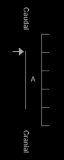
Tomosynthesis indicator Shown in the viewport for breast tomosynthesis studies to show the location of the current slice in relation to the other images in the series. The indicator shows directional markers to indicate whether you are scrolling in a caudal / cranial, or a medial / lateral direction.
|
Mammography base view
Warning
When the breast zoom to fit feature is applied on a series basis, the resulting zoom to fit action on the images with different orientation may zoom the anatomy of any of the images out of the field of view.
To avoid this, create a hanging protocol to split out the images of the series into single series so each series consists of a single orientation.
When a mammography study is first opened, eUnity automatically presents a consistent layout for evaluation and interpretation. This default layout is referred to as base view. eUnity will not apply the base view to mammography studies if the following required DICOM attributes are missing, and the Orientation Marker is set as an Unknown View.
- Patient Orientation (0020,0020)
- Image Laterality (0020,0062)
- View Code Sequence (0054,0220)
- View Code Modifier Sequence (0054,0222), when present
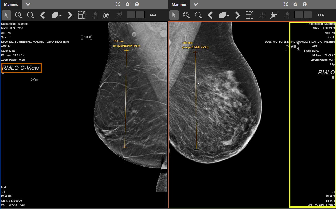
Figure 4: : eUnity mammography base view
|
Ventral view The viewer displays the left breast with the nipple pointing to the right of the viewport and a right breast displays the nipple pointing to the left of the viewport. |
|
Chest wall alignment The chest walls are aligned to the viewport edge. |
|
Same size Images are scaled in the viewport so that images from the same patient, performed at different times on different detectors can be displayed at the same size. This allows for evaluation of developing densities and allows the radiologist to evaluate for a change in size of known lesions during temporal comparison with prior digital images or even film mammograms. See the yellow measurement lines in the above image. |
|
Demographic overlay The viewer shows the demographic overlay opposite the chest wall by default so it does not cover the relevant anatomy. The demographic overlay is outlined in yellow in the above image. |
|
Orientation marker The orientation marker is always shown opposite the chest wall and it cannot be turned off. If C-View (Hologic : Synthesized 2D Mammographic Imaging) is present, it is added to the orientation marker so that the user is always aware that they are viewing a synthesized image (for example, RCC C-View). The orientation marker is outlined in orange in the above image. |
|
Flip images eUnity always flips images that would otherwise present upside down. When an image is flipped, the viewer shows the rotate indicator in the viewport. |
|
Breast tomosynthesis viewing eUnity shows a breast tomosynthesis indicator in the viewport to show the type of breast tomosynthesis and location of the current slice in relation to the other images in the series. |
Air gap suppression
eUnity uses air gap suppression so that the air gap area (the viewport background) retains its color when the Window Level is changed or an image is inverted in a mammography study.
For air gap suppression to work, the image cannot be a secondary capture, color, or lossy and the Pixel Padding Value (0028,0120) and Pixel Padding Range Limit (0028,0121) tags must be populated in the DICOM header. If these values are missing or incorrect, air gap suppression may not work as expected. Note that air gap supression can be disabled by a system administrator.